Fds And Fdp Anatomy - Flexor Tendon Injury Musculoskeletal Medicine For Medical Students Orthopaedicsone / Overlying these are three muscles, flexor carpi ulnaris (fcu), palmaris longus and flexor carpi radialis (fcr) (fig.
Get link
Facebook
X
Pinterest
Email
Other Apps
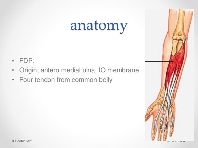
Fds And Fdp Anatomy - Flexor Tendon Injury Musculoskeletal Medicine For Medical Students Orthopaedicsone / Overlying these are three muscles, flexor carpi ulnaris (fcu), palmaris longus and flexor carpi radialis (fcr) (fig.. The fds tendons are the most palmar, and the fds tendons to the long and ring fingers are most superficial. It can sometimes be classed as a superficial muscle, but in most individuals, it lies between the deep and superficial muscle layers. Normal flexor digitorum profundus (fdp) and superficialis tendons (fds) at the level of the metacarpal neck. The flexor digitorum profundus muscle is found in the deep layer of the anterior forearm. (see finger and thumb anatomy.) the flexor digitorum profundus (fdp) tendon travels along the volar side of the palm and finger and passes distally through a split in the flexor digitorum superficialis (fds) tendon to insert at the base of the distal phalanx (figure 1 and figure 2).
The flexor digitorum profundus lies directly beneath the flexor digitorum superficialis. The muscle fans out into four tendons (one to each of the second to fifth fingers) to the palmar base of the distal phalanx. The flexor digitorum profundus is supplied by the anterior interosseous artery, which is a branch of the common interosseous artery. Anatomy of the flexor tendons. The fdp tendon is not present.
Testing Tendons Occupational Therapy Hand Therapy Massage Therapy from i.pinimg.com Flexor zone ii is defined as the region spanning the proximal aspect of the a1 pulley to the insertion of the flexor digitorum superficialis (fds) tendon. It can sometimes be classed as a superficial muscle, but in most individuals, it lies between the deep and superficial muscle layers. Along with the flexor pollicis longus and pronator quadratus muscles, it comprises the deep flexor compartment of the forearm. Overlying these are three muscles, flexor carpi ulnaris (fcu), palmaris longus and flexor carpi radialis (fcr) (fig. The two tendinous septa (ts) were found: The flexor digitorum profundus is situated on the ulnar side of the forearm, immediately beneath the superficial flexors. This definition incorporates text from a public domain edition of gray's anatomy (20th u.s. The fds is detached from the middle phalanx and advanced to the distal fdp stump;
Ebraheim's educational animated video describes the anatomy of the flexor digitorum profundus muscle.the flexor digitorum profondus is a muscle in the fo.
The key features of this technique are: (see finger and thumb anatomy.) the flexor digitorum profundus (fdp) tendon travels along the volar side of the palm and finger and passes distally through a split in the flexor digitorum superficialis (fds) tendon to insert at the base of the distal phalanx (figure 1 and figure 2). Musculus flexor digitorum profundus) is a flat muscle of the forearm that belongs to the anterior muscle group and is situated in the third or deep layer. Note the relationship between the flexor digitorum superficialis (fds) and flexor digitorum profundus (fdp) tendons.c, extensor tendon compartments. This definition incorporates text from a public domain edition of gray's anatomy (20th u.s. It can sometimes be classed as a superficial muscle, but in most individuals, it lies between the deep and superficial muscle layers. The flexor digitorum superficialis is the only muscle of the intermediate compartment. And the proximal stump of the fdp is advanced to reestablish proper lumbrical tension and sewn to the fds tendon proximally. The fds is detached from the middle phalanx and advanced to the distal fdp stump; (a) drawing (lateral view) shows the fdp and fds tendons and their points of insertion. Or it may be replaced by a small fusiform muscle arising from the long flexor tendon or from the quadratus plantae. The fdp originates from the anterior and medial surfaces of the ulna, runs deep to the tendons of fds, passes through the carpal canal and then perforates the tendons of fds to insert to the base. The fdp tendon is not present.
Flexor tendon anatomy the flexor tendons of the digits enter the carpal tunnel in a generally consistent anatomic relationship. The muscle that moves these tendons is a common muscle belly shared by all the fingers. The treatment of zone i flexor digitorum profundus (fdp) avulsions and lacerations requires reattachment of the tendon to the distal phalanx. The key features of this technique are: The flexor digitorum profundus lies directly beneath the flexor digitorum superficialis.
Flexor Tendon Injuries from image.slidesharecdn.com The muscle fans out into four tendons (one to each of the second to fifth fingers) to the palmar base of the distal phalanx. The fds is detached from the middle phalanx and advanced to the distal fdp stump; The flexor digitorum profundus is situated on the ulnar side of the forearm, immediately beneath the superficial flexors. Edition of gray's anatomy of the human body, published in. The flexor digitorum superficialis is the only muscle of the intermediate compartment. Each fds tendon inserts on the volar surface of the mid diaphysis of p2 as two separate slips. Similarly, each fdp tendon inserts on the volar aspect of the proximal diaphysis of p3. (b) photograph of a dissected anatomic specimen shows a superficial volar view of the chiasm of the fds tendon.
The fds is detached from the middle phalanx and advanced to the distal fdp stump;
Or it may be replaced by a small fusiform muscle arising from the long flexor tendon or from the quadratus plantae. This definition incorporates text from a public domain edition of gray's anatomy (20th u.s. Overlying these are three muscles, flexor carpi ulnaris (fcu), palmaris longus and flexor carpi radialis (fcr) (fig. The fdp tendon is not present. The fdp and fpl tendons are found in the deepest level of the carpal tunnel. The muscle belly divides into 4 tendons. Flexor zone ii is defined as the region spanning the proximal aspect of the a1 pulley to the insertion of the flexor digitorum superficialis (fds) tendon. It is considered an extrinsic hand muscle because it acts on the hand while its muscle belly is located in the forearm. The muscle fans out into four tendons (one to each of the second to fifth fingers) to the palmar base of the distal phalanx. Flexor digitorum superficialis flexor carpi ulnaris deep flexors. The flexor digitorum profundus is supplied by the anterior interosseous artery, which is a branch of the common interosseous artery. (a) drawing (lateral view) shows the fdp and fds tendons and their points of insertion. Flexor digitorum superficialis (musculus flexor digitorum superficialis) flexor digitorum superficialis is the largest muscle of the anterior compartment of the forearm.
(b) photograph of a dissected anatomic specimen shows a superficial volar view of the chiasm of the fds tendon. A consistent dense palmar and dorsal vascular supply to the tendon at its insertion into the distal phalanx was observed based on sources from both. Musculus flexor digitorum profundus) is a flat muscle of the forearm that belongs to the anterior muscle group and is situated in the third or deep layer. Flexor digitorum superficialis (musculus flexor digitorum superficialis) flexor digitorum superficialis is the largest muscle of the anterior compartment of the forearm. Edition of gray's anatomy of the human body, published in.
An Anomaly Of Flexor Muscles Of The Fifth Little Finger Of The Hand An Anatomical Case Report from www.scielo.br The anterior interosseous artery is accompanied by the palmar interosseous branch of the median nerve and gives off muscular branches to the flexor digitorum profundus and flexor pollicis longus muscles. Flexor tendon anatomy the flexor tendons of the digits enter the carpal tunnel in a generally consistent anatomic relationship. The fdp tendon is not present. Ebraheim's educational animated video describes the anatomy of the flexor digitorum profundus muscle.the flexor digitorum profondus is a muscle in the fo. (a) drawing (lateral view) shows the fdp and fds tendons and their points of insertion. A consistent dense palmar and dorsal vascular supply to the tendon at its insertion into the distal phalanx was observed based on sources from both. Overlying these are three muscles, flexor carpi ulnaris (fcu), palmaris longus and flexor carpi radialis (fcr) (fig. The muscle belly divides into 4 tendons.
(b) photograph of a dissected anatomic specimen shows a superficial volar view of the chiasm of the fds tendon.
Similarly, each fdp tendon inserts on the volar aspect of the proximal diaphysis of p3. The fds tendons are the most palmar, and the fds tendons to the long and ring fingers are most superficial. Flexor digitorum superficialis (fds), also known as flexor digitorum sublimis, is a muscle in the second (intermediate) layer of the anterior compartment of the forearm. The two tendinous septa (ts) were found: The muscle belly divides into 4 tendons. The fds is detached from the middle phalanx and advanced to the distal fdp stump; Anatomy of the flexor tendons. Edition of gray's anatomy of the human body, published in. The anterior interosseous artery is accompanied by the palmar interosseous branch of the median nerve and gives off muscular branches to the flexor digitorum profundus and flexor pollicis longus muscles. (b) photograph of a dissected anatomic specimen shows a superficial volar view of the chiasm of the fds tendon. The fds tendons insert on the middle phalanx (p2), whereas the fdp tendons insert on the distal phalanx (p3) 1, 2 . This muscle extends from the proximal part of the ulna to the distal phalanges of the 2nd to 5th digit. This definition incorporates text from a public domain edition of gray's anatomy (20th u.s.
(see finger and thumb anatomy) the flexor digitorum profundus (fdp) tendon travels along the volar side of the palm and finger and passes distally through a split in the flexor digitorum superficialis (fds) tendon to insert at the base of the distal phalanx (figure 1 and figure 2) fdp anatomy. The vascular anatomy of the flexor digitorum profundus (fdp) tendon insertion is described by using a vascular injection and modified spalteholtz tissue clearing protocol in 36 human cadaver digits.


Comments
Post a Comment